What is it?
The anterior cruciate ligament (ACL) is one of the primary ligaments in the knee, connecting the femur (thigh bone) to the tibia (shin bone). It is responsible for preventing the tibia from sliding forward, and it provides rotational stability to the knee. ACL injuries commonly occur during athletics, especially in cutting and pivoting sports, such as football, soccer, and basketball.
WHAT ARE SIGNS AND SYMPTOMS OF AN ACL INJURY?
Common symptoms of rotator cuff tears include:
• The knee suddenly giving out during a non-contact activity
• An audible pop
• Pain and swelling
• Difficulty walking
Diagnosis
In the office, a complete history and physical exam is obtained. The knee is examined for any deformity or tenderness to palpation. A comprehensive test of all the ligaments is performed and oftentimes compared to the uninjured side. Imaging studies, such as an X-Ray and MRI, may be necessary to confirm the diagnosis and identify associated injuries, such as meniscal tears.
Treatment
Conservative treatment of ACL injuries is an option in elderly patients or patients who do not participate in cutting activities. Such treatments include rest, ice, anti-inflammatories, and physical therapy. Crutches and a knee brace may be used initially to support the injured knee.
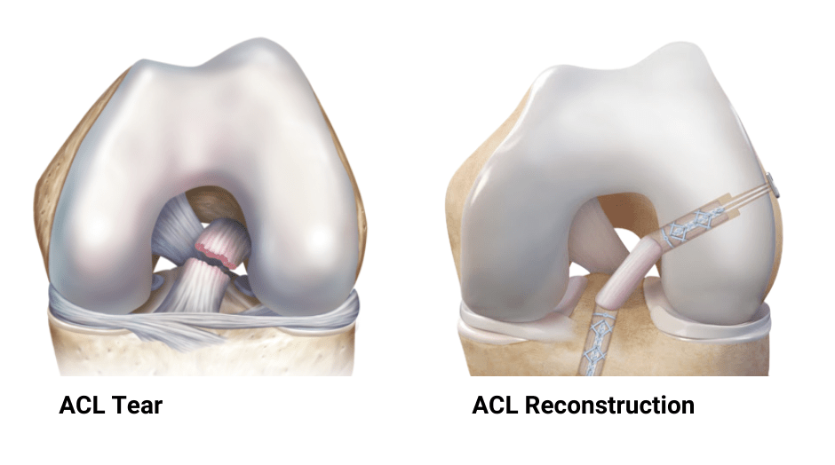
Surgical treatment of ACL injuries is generally recommended in active individuals who wish to return to their sport or activity. In most cases, the injured ligament cannot simply be repaired. ACL reconstruction involves placing a new ligament (graft) into the knee, which, over time, matures into the new ACL. This is typically done in an outpatient setting under regional anesthesia, allowing patients to return home the same day.
ACL graft choices can come from the patient (autograft) or from a cadaver (allograft). Multiple studies have demonstrated that autografts, especially in younger patients, are associated with superior outcomes and lower failure rates. The most common autografts used include the patellar tendon, hamstring tendon, and quadriceps tendon. Each graft has their own advantages and disadvantages and it is important for the patient and physician to discuss them in detail in order for the patient to make an informed decision.
In conjunction with supervised physical therapy, ACL reconstruction has been shown to support the patient’s successful return to their sport.
RECOVERY
Following surgery, patients are placed in a long hinged knee brace for six weeks and are allowed to bear weight immediately (except in cases where there is a combined meniscus root repair or cartilage repair). Physical therapy begins within the first week of surgery, with emphasis on knee range of motion. At six weeks, the brace is removed and the focus turns to gait training and strengthening. A jogging program is initiated about 3-4 months postoperatively, followed by an agility program at 5-6 months. Sport specific drills are initiated at six months and patients generally return back to sports around 9-12 months postoperatively.
ARTHROSCOPIC ACL RECONSTRUCTION
The following animation demonstrates an all-inside arthroscopic reconstruction of the ACL with either a hamstring or quadriceps tendon graft. Following graft harvest and preparation, small sockets are created in both the femur and tibia. The graft is then introduced into the femoral socket and then into the tibial socket. Graft fixation is performed by tensioning and tying the sutures over a button.